|
|
click photo for larger file

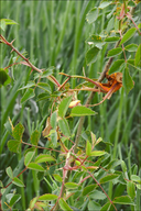
Phragmidium mucronatum
Rose Rust
|
Photographer: Dr. Amadej Trnkoczy
ID: 0000 0000 0517 0453 (2017-05-15)Copyright © 2017 Dr. Amadej Trnkoczy
|
|
INFORMATION PROVIDED WITH THE PHOTO
|
date of photo Apr 24, 2017
latitude 45.08710 longitude 14.44961
View on Google Maps.
location
West part of Krk Island, between villages Brzec and Poljica, Adriatic Sea (Kvarner bay, Croatia)notes Slo.: rja vrtnic - syn.: Puccinia mucronata Pers., Phragmidium rosae (Pers.) Rostr., and more than 50 other names (Index Fungorum)! - Habitat: (sub)mediterranean maquis scrubland; narrow, dirt road side, almost flat terrain, calcareous, skeletal ground, Karst region; warm, dry and quite sunny place; average precipitations ~ 900 mm/year, average temperature 12-14 deg C, elevation 125 m (410 feet), (sub)mediterranean phytogeographical region. Substratum: small, living shrub of Rosa sp. (canina ?) leaves and buds. Comments: Phragmidium mucronatum is a common rust fungus growing on roses (Rosa). It feeds off the plant’s own supplies thereby weakening it. This is the reason why gardeners hate it. Taxonomy of rust fungi is very 'difficult' (see number of synonym names for Phragmidium mucronatumit above!). Also its life cycle is very complex. Leaf rust of roses grows through several different stages. It produces four different forms of spores. The original pustules (aecia) produce aecidospores (each spore has two nuclei, they appear usually in chains), which infect other leaves or other parts of the plant and in turn produce the yellow-orange pustules and wounds. They form urediniospores (dikaryotic spores, which disperse widely, spreading infection), which germinate to form black pustules. They contain teliospores (thick-walled resting spores) which overwinter on fallen leaves, to germinate in the spring producing basidiospores (spores produced by specialized fungal cells called basidia), which are blown by wind. Pictures show the orange, spring time stage with urediospores. Urediospores not smooth, minutely, (sharply ?) warted; spores of irregular shape. Dimensions: 20 [24.3 ; 26.2] 30.5 x 15.3 [19.9 ; 21.9] 26.5 microns; Q = 0.9 [1.2 ; 1.3] 1.5; N = 30; C = 95%; Me = 25.2 x 20.9 microns ; Qe = 1.2. Olympus CH20, NEA 40x/0.65, magnification 400x (spores), NEA 10x/0.25, magnification 100x (squash), dry material; in water. AmScope MA500 digital camera. Herbarium: Mycotheca and lichen herbarium (LJU-Li) of Slovenian Forestry Institute, Večna pot 2, Ljubljana, Index Herbariorum LJF Ref.: (1) http://eol.org/pages/1009444/hierarchy_entries/51043771/media (2) http://www.wikiwand.com/en/Phragmidium (3) http://www.inaturalist.org/taxa/383622-Phragmidium-mucronatumcamera Nikon D700 / Nikkor Micro 105mm/f2.8
contributor's ID # Bot_1049/2017_DSC7443 photo category: Fungi - fungi
|
MORE INFORMATION ABOUT THIS FUNGUS
|
| common names
Rose Rust, Common Leaf Rust (photographer)
View all photos in CalPhotos of Phragmidium mucronatum Check Google Images for Phragmidium mucronatum |
|
The photographer's identification Phragmidium mucronatum has not been reviewed. Click here to review or comment on the identification. |
|
Using this photo The thumbnail photo (128x192 pixels) on this page may be freely used for personal or academic purposes without prior permission under the Fair Use provisions of US copyright law as long as the photo is clearly credited with © 2017 Dr. Amadej Trnkoczy.
For other uses, or if you have questions, contact Dr. Amadej Trnkoczy amadej.trnkoczy[AT]siol.net. (Replace the [AT] with the @ symbol before sending an email.) |
|
|
