|
|
click photo for larger file

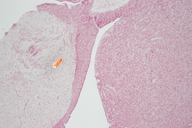
Pituitary (hypophysis), 10x
Felis silvestris catus
|
Photographer: Sarah Werning
ID: 0000 0000 0807 0873 (2007-08-15)Copyright © 2007 Sarah Werning
|
|
INFORMATION PROVIDED WITH THE PHOTO
|
date of photo Aug 5, 2007
notes Pituitary (hypophysis), H&E stain. Taken at 10x. This is IB 131L slide #51. This image shows the anterior pituitary (= adenohypophysis; darkly stained, on the right in this image), the posterior pituitary (= neurohypophysis; lightly stained, on the left in this image), and the inferior portion of the infundibulum (= infundibular stalk or pituitary stalk). The pars intermedia is part of the anterior pituitary, but appears as a dark band on the anterior (right in this image) side of the posterior pituitary. The anterior pituitary is densely cellular and converts hormone precursors made in the hypothalamus into hormones, which the anterior pituitary also stores and releases. The posterior putuitary is primarily composed of axons that travel down from cell bodies located in the hypothalamus. The hypothalamus produces hormones and sends them down axons traveling through the infundibulum to the posterior pituitary. These hormones are stored in the posterior pituitary until they are released.camera Nikon D70s, original resolution: 300dpi, 3008x2000pxls at 10x magnification
photo category: Misc. - histology
|
MORE INFORMATION
|
| View all photos in CalPhotos of Felis silvestris catus Check Google Images for Felis silvestris catus |
| |
|
Using this photo The thumbnail photo (128x192 pixels) on this page may be freely used for personal or academic purposes without prior permission under the Fair Use provisions of US copyright law as long as the photo is clearly credited with © 2007 Sarah Werning.
For other uses, or if you have questions, contact Sarah Werning swerning[AT]gmail.com. (Replace the [AT] with the @ symbol before sending an email.) |
|
|
