|
|
click photo for larger file

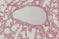
Lung alveoli and vein, 25x
Cavia porcellus Guinea Pig
|
Photographer: Sarah Werning
ID: 0000 0000 0707 1490 (2007-07-27)Copyright © 2007 Sarah Werning
|
|
INFORMATION PROVIDED WITH THE PHOTO
|
date of photo Jul 9, 2007
notes Thin section of alveoli of the lung showing a vein, Mallory-Azan stain. Taken at 25x. IB 131L slide #31. The alveoli are lined with simple squamous epithelium. The alveoli are small sacs or hollow balls of simple squamous epithelium. In this image, the thinner tunica media is stained pink and the thicker tunica externa is stained blue. A faint spread of erythrocytes (red blood cells) can be seen across the lumen. Note the irregular shape of this vein.camera Nikon D70, original resolution: 300dpi, 3008x2000pxls at 25x magnification
photo category: Misc. - histology
|
MORE INFORMATION
|
| common names
Slide #31 - Lung, Alveoli, Vein, 25x
Guinea Pig (photographer)
View all photos in CalPhotos of Cavia porcellus Check Google Images for Cavia porcellus |
| |
|
Using this photo The thumbnail photo (128x192 pixels) on this page may be freely used for personal or academic purposes without prior permission under the Fair Use provisions of US copyright law as long as the photo is clearly credited with © 2007 Sarah Werning.
For other uses, or if you have questions, contact Sarah Werning swerning[AT]gmail.com. (Replace the [AT] with the @ symbol before sending an email.) |
|
|
